Thursday 11 December 2014

Nic Tapon: Growth Control by the Hippo Pathway

Nic Tapon is one of the few scientists who have made major contributions to the discovery of a new signal transduction pathway. As a result of tumour suppressor screens in the fruit fly he and others identified a set of genes/proteins that regulate development, organ growth/size, stem cell function and whose dysregulation is involved in the development of many types of cancer. This regulatory system is now known as the Hippo pathway. 
Nic Tapon & fruit fly, the key model of his research

Recent research has connected Hippo members to many other regulatory systems including AMPK and mTOR (a hint to exercise physiologists) and Nic's team has contributed important discoveries. When giving a talk at Aberdeen Nic was willing to answer some questions related to his research. 


1) How did you become involved in the tumour suppressor screens and why did you use the fruit fly as a model?
My PhD was a cell culture-based project, but when I wrote the introduction to my thesis, I became fascinated by the fly and worm genetics literature and the power of screens. The possibility of doing complex genetic manipulations very rapidly was also part of the attraction. I was lucky to be offered a postdoc position in Iswar Hariharan’s lab at a time when genetic mosaicism (the ability to genetically manipulate small patches of tissue in an otherwise wild type organism) was revolutionising fly genetics. This created a gold rush of phenotype-based screens to study all sorts of processes, from tumour suppression to neurodegeneration. At the same time, the fly genome was being sequenced, which made mapping the mutations much easier. The discovery of the Hippo pathway was part of this genetic gold rush. These were exciting times!

2) When and how did you note that the tumour suppressor genes were not independent genes but constitute a pathway?
During the course of my screen, I identified mutations in two genetic loci with similar phenotypes, salvador and dali (long story…). So it was the similarity between the two phenotypes (more proliferation, less apoptosis) that told us these two genes might function in a common pathway. dali turned out to be the previously identified tumour suppressor kinase warts/lats, which had been discovered by Peter Bryant and Tian Xu. Marius Sudol had identified the WW domain as a small protein-protein interaction motif, and Salvador has two WW domains, while Warts has several WW domain-binding motif. Kieran Harvey then showed that Salvador and Warts can physically interact in vitro, and we went from two orphan tumour suppressors to a (modest) pathway!

3) How did people in the field agree on the term ‘Hippo’?
Georg Halder had called his mutants hippo because the head of mutant flies had a puckered appearance reminiscent of hippopotamus skin. This seemed more fun than Drosophila Mammalian Sterile 20-like, so the rest of us adopted that name! DJ Pan started to use the term “Hippo pathway” in his 2003 Cell paper (Wu et al), and the rest is history…

4) Why do you think was the Hippo pathway was overlooked for so long despite its ubiquitous expression and importance?
The core kinase cassette was actually discovered in yeast as the Mitotic Exit Network/Septation Initiation Network before the Hippo pathway was identified in flies. The connection with tissue growth was made thanks to advances in genetic mosaicism, which allowed phenotype-based screens for tumour suppressor genes. So I think it was technological advances that were needed to kick-start the field.

5) The Tead transcription factors, which are now seen as the key downstream regulators of the pathway, were first characterised often in the context of skeletal muscle. Were you surprised by this (Hippo was mainly studied in epithelial cells) and how did this affect your thinking about the Hippo pathway?
All major signalling pathways are used reiteratively in many processes. Although it was initially identified in the context of growth control, the Hippo pathway is no different. So no, not surprised.

6) What are your next (non-confidential) research plans?
That would be telling! Understanding the wildtype function of the Hippo pathway (see Lawrence and Casal, Dev Biol 2013).

Friday 16 May 2014

First Aberdeen Myology Meeting, 25.4.2014

On the 25th of April, 2014 we organised the First Aberdeen Myology Meeting with support from the MRC grant that is shared between Pete Zammit, Cosimo De Bari and Henning Wackerahge. For the meeting the speakers were:

Prof. Pete Zammit, King's College London
Prof. Mike Rennie, University of Nottingham
Prof. Steve Harridge, King's College London
Dr. Henning Wackerhage, University of Aberdeen

In order to make it suitable for a wider audience we asked the speaker to give the first half of the talk in a way which is suitable for the general public followed by a second half which is specific to their research. 

Pete Zammit started the afternoon with a basic introduction to skeletal muscle and genetic disorders which worked very well as it reminded the muscle researchers of some basic facts (human muscle fibres can be up to 20 cm long and probably have tens of thousands of nuclei) and at the same time introduced the non-muscle researchers and general public to the topic. 

He then went on to talk about his work on his teams work on laminopathies. These are genetic diseases involving mutations in genes that encode proteins which are in the nuclear lamina and in some cases cause muscular dystrophy. He also presented data on exon skipping, a promising treatment to turn dysfunctional genes into genes that result in a moderately affected protein and thereby result in treatment. This is promising research as exon skipping has been tried with success in patients with muscular dystrophy. 

Figure 1. Prof. Pete Zammit during his talk. 

Mike Rennie gave a talk introducing sarcopenia, the loss of muscle mass during normal ageing, and the concept of 'anabolic resistance' which refers to the observation that older human muscles respond with less protein synthesis to anabolic stimuli such as amino acids than young muscle. Mike has led human muscle protein synthesis in the field and thus it was an excellent review of the work which spanned several decades. Towards the end he has presented a reinterpretation of his data which he is currently presenting at various meetings.

Steve Harridge started by introducing Masters athletes and his studies on Masters athletes as a model for ageing. The world records of Masters athletes show that at least some individuals can maintain a very high level or aerobic capacity and strength into old age. He then presented work by Chibeza Agley who was an oustanding with Steve and who is now a Postdoc in Cambridge: 
http://www.stemcells.cam.ac.uk/researchers/researchersbygroup/silvalabmembers/chibeza-agley
Bezza, as he is known, optimised the methods for isolating myoblasts and fibroblasts from human muscle biopsies and then differentiated these cells. This research showed that fibroblasts but not myoblasts can differentiate into fat cells: http://jcs.biologists.org/content/early/2013/09/23/jcs.132563.abstract

 Figure 2. Prof. Steve Harridge during his talk

At the end I introduced our research on the Hippo pathway in rhabdomyosarcoma which just got accepted by Cancer Cell. This work was led by Fernando Camargo and Annie Tremblay at Harvard who did the mouse models whilst we worked on human cancer cells and stained human tissues. Edoardo Missiaglia, Pete Zammit and Janet Shipleys were other collaborators on this large scale project. 

In the first part I was introducing the hallmarks of cancer (sustained proliferation etc) and how mutations in cancer cells or the germline drive these hallmarks. In the second part I presented the data showing that started with the discovery that the Hippo protein Yap can drive the proliferation of satellite cells. We then asked the question whether persistent Yap hyperactivity would lead to cancer and found that it did but only in activated but not quiescent satellite cells. These cancers in mice mimicked human embryonal rhabdomyosarcoma (ERMS), which is the most common form of this cancer. When Yap is reduced in human ERMS cells then these cells differentiate into normal, differentiated muscle tissue. We also studied the molecular mechanisms by which Yap alone can drive this cancer and many other aspects. This will be covered in another blog entry. 
Figure 3. Dr. Henning Wackerhage during his talk on Cancer and Muscle. 

So overall it was good to see many colleagues, students and researchers attending the myology day. I think the combination of an introductory part and an actual research part worked well, giving non-muscle research participants the chance to learn.

Last but not least the acknowledgements. Thanks to all the speakers for travelling to Aberdeen! Thanks to Rosa, Claire, Vanessa, Roby, Abdalla and Raphael for organising the day which involved advertisment, communication, catering, transport, accommodation and so on. You have done a fantastic job!
VDM, AM, HW

Wednesday 8 January 2014

Scottish Hippo workshops

The annual Scottish Hippo Workshop
The Hippo pathway is a recently identified, ubiquitously expressed signal transduction pathway which has major functions during development, in stem cells, organ size regulation and cancer. In Scotland, groups at the Universities of St. Andrews, Edinburgh and Aberdeen have started to work on this pathway. To foster collaboration we have invited each other for talks (Dr Marius Sudol and Prof Junichi Sadoshima gave talks in Scotland) and we have started to organize Scottish Hippo workshops where we combine Hippo talks with a joint meal afterwards. The aim of this blog entry is to briefly introduce the Scottish Hippo scene and the Scottish Hippo workshops.

First Scottish Hippo workshop, University of Aberdeen, 5.12.2012
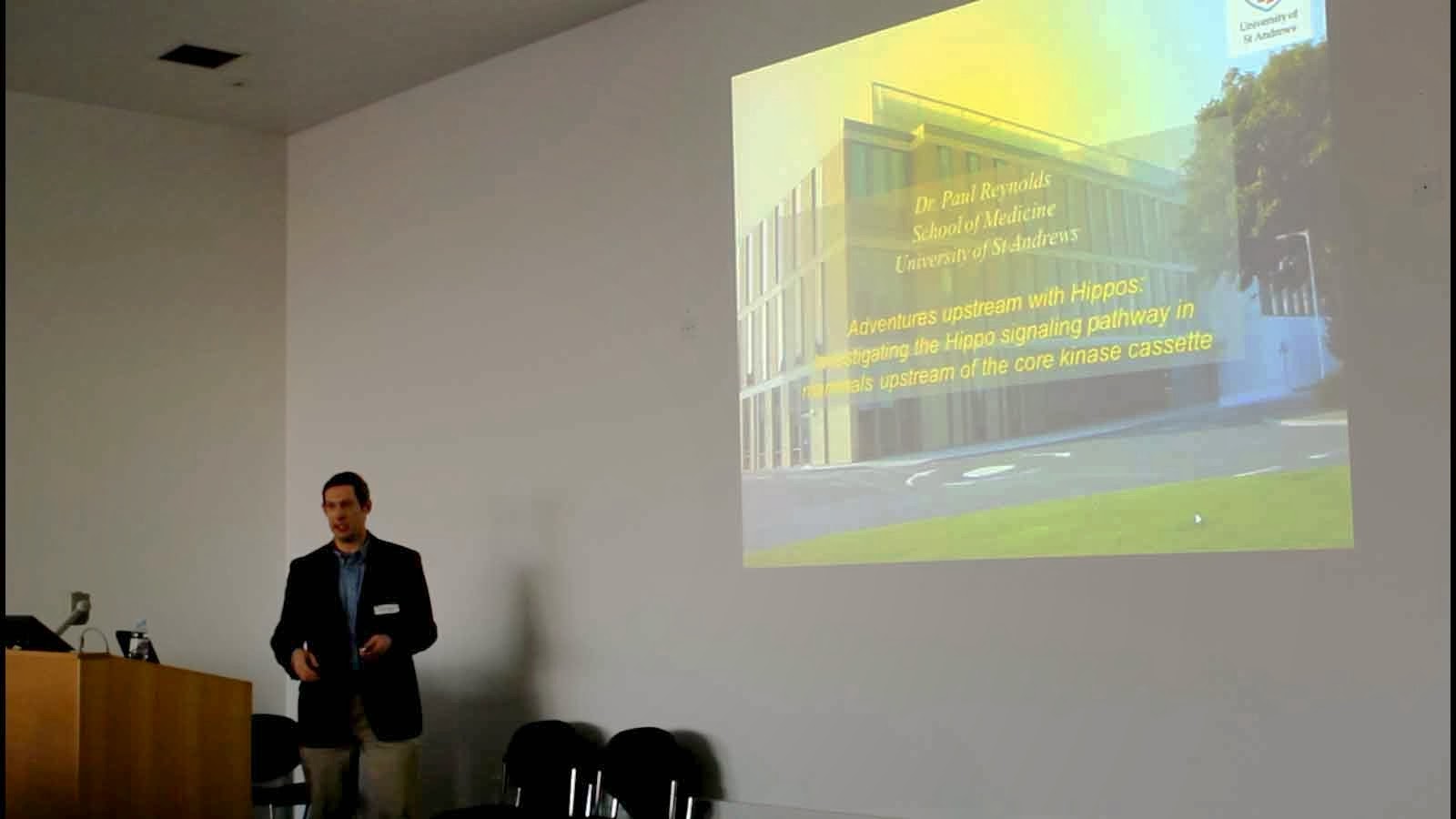
The keynote speaker during the first workshop was Dr. Paul Reynolds from the University of St. Andrews and his talk was entitled ‘Adventures upstream with Hippos: investigating the Hippo pathway in mammals upstream of the core kinase cassette’. It featured the role of Willin/Frmd6 and Kibra upstream in the Hippo pathway which are major contributions by the St. Andrews team to the pathway architecture and the role of the Hippo pathway in breast cancer (Figure 1).
Figure 1 Paul Reynolds speaking at the Institute of Medical Sciences, Aberdeen December 2012.

Second Scottish Hippo Workshop, University of St Andrews, 13.12.2013
The second Scottish Hippo Workshop was held at the University of St Andrews and organised by Dr. Paul Reynolds. The conference was kick started by Professor Frank Gunn-Moore, where he spoke about the involvement of the Hippo Pathway in Neuroscience. The conference finished with a wrap up by Dr Henning Wackerhage from the University of Aberdeen who gave a talk entitled ‘When Hippo met MCAT: How Hippo links to stem cells, cancer and dystrophy in skeletal muscle’. Dr. Anke Roelofs gave a talk on Hippo in cartilage followed by shorter PhD student talks.
Figure 2 Frank Gunn-Moore giving his talk on Willin/FRMD6 and the peripheral nervous system

Figure 3 Henning Wackerhage talking on ‘When Hippo met MCAT. How Hippo links to stem cells, cancer and dystrophy in skeletal muscle’.

So if you are interested in joining the Scottish Hippo scene then please e-mail Henning (h.wackerhage@abdn.ac.uk) and we will keep you in the loop.

Vanessa De Mello (PhD student at Aberdeen with an interest in science journalism)
Henning Wackerhage

Tuesday 12 November 2013

How to subclone

What is subcloning and what is it used for?
A common method to study the function of a gene is either to knock it out or to overexpress it (knock in). This can be done in mice or in cultured cells. Exercise physiologists use this method for example to test whether a potential regulator of adaptive responses does indeed trigger the hypothesized adaptations when its concentration is increased.

In many cases the DNA constructs can be obtained from colleagues. Alternatively many DNA constructs can be purchased from Addgene (www.addgene.org). However, not all vectors are suitable for all cells and sometimes you wish to add your own gene into a vector in order to study it. For this DNA need to be cut out a longer piece of DNA or cDNA and inserted into a suitable vector. This process is termed subcloning.

In this blog entry Abdalla Diaai (Figure 1), who is a PhD student in our group, explains how to subclone and answers some key questions about subcloning. Abdalla has recently subcloned several Hippo gene inserts into a vector that is suitable for the use in satellite cells. Abdalla and Dr. Roby Urcia, a post doc in our group, are the cloning experts in our team.

Figure 1. Abdalla in the DNA lab of the Musculoskeletal Group in Aberdeen.

Over to Abdalla: OK, In figure 2 I have drawn a schematical overview which illustrates how a DNA insert (shown in red) is taken out of a donor plasmid and inserted into a recipient plasmid. Plasmids are pieces of circular DNA which can be inserted into cells as vectors for DNA inserts that may encode a gene knock in (overexpression) or knock out construct.
Figure 2.In the first step the DNA of interest (red) in the donor plasmid needs to be cut out. The molecular scissors are termed restriction enzymes and they cut DNA at specific, short DNA sequences. In this examples no suitable restriction sites are found near the gene of interest so we are introducing EcoRI and NotI (these sites are cut by enzymes termed EcoRI and NotI) sites upstream and downstream of the insert so that we can cut it out. These restriction sites are added by PCR using unique primers which contrain the EcoRI and NotI restriction sites respectively. After that the DNA of interest is cut out by adding the EcoRI and NotI restriction enzymes. We also use the same enzymes to open up the recipient plasmid which already has these sites and then ligate our gene of interest into the recipient plasmid to yield the subcloned plasmid. Easy! 

As with many molecular biology method quite a bit of know how is required to successfully sub clone. Here I will answer a few key questions.

Question: How do I cut DNA?
Answer: For this you can use restriction enzymes which cut DNA at specific DNA sites. In Figure 2 EcoRI and NotI are restriction enzymes which cut DNA at EcoRI and NotI restriction sites. Restriction enzymes are the scissors of molecular biologists and restrictions sites are the specific, short DNA motifs, which are cut by these scissors.

Question: How do you find restriction enzymes in a DNA sequence?
Answer: Several websites are available to identify the short sites in a DNA sequence where specific restriction enzymes cut. I am using Serial Cloner which is a free download (http://serial-cloner.en.softonic.com/) and after trying it a bit you should be able to identify restriction sites.

Question: The Serial Cloner programme comes up with many restriction sites. Which ones should I use?
Answer: When selecting restriction sites to cut out your gene of interest the key criteria are:
-       Restriction sites must lie outside the insert (gene of interest);
-       The restriction sites need to be in the desired location in your recipient plasmid (plasmids often have multiple cloning sites (abbreviated as ‘MCS’) with several restriction sites). However, they should not be elsewhere in your recipient plasmid or you will cut it where you do not want to cut it.
-       Ideally choose restriction enzymes that work in the same restriction enzyme buffer as this makes things easier.
-       Try to identify two different restriction sites and enzymes for your subcloning because otherwise the recipient plasmid might self-ligate (i.e. form a ring of DNA without the gene of interest or insert in it!). You can get around that problem by using a phosphatase like Shrimp Alkaline Phosphatase which catalyzes the dephosphorylation of 5´ and 3´ ends of DNA. Such dephosphorylation prevents religation of linearized plasmid DNA.

If you cannot find enzymes that meet these criteria then there are alternatives such as:
-      You can modify the MCS of your recipient plasmid and add new restriction sites by using ‘annealed-oligos’ which enable you to add short stretches of DNA into your recipient plasmid. More information can be found on (http://www.addgene.org/plasmid_protocols/annealed_oligo_cloning/).
-      Use the annelead oligos as primers and PCR to add restriction sites to the MCS of your recipient plasmid.

How to run the PCR to amplify your construct and how to purify the PCR product?
In order to have sufficient insert DNA (gene of interest) you will need to run a PCR to amplify your insert DNA. For this it is important to use a high fidelity taq polymerase (Platinum® Pfx DNA Polymerase-Invitrogen) to minimize the number of errors. The longer the expected PCR product is, the more important the fidelity of the polymerase becomes. You should select an annealing temperature based on the melting temperature (Tm) of the portion of the primer that binds to the sequence to be amplified (the hypridization sequence), not the Tm of the entire primer. After the PCR reaction is complete, isolate your PCR product from the rest of the PCR reaction using a kit, such as the QIAquick PCR Purification Kit (Qiagen). The PCR product is now ready for restriction digestion. If you amplify your PCR product from a plasmid, it’s advisable to digest your PCR product in DpnI as this digests methylated DNA. It is important to run the product on a gel. This allows you to visualize that your PCR product is the anticipated size and that your band is strong (indicating that the PCR reaction worked and that you have a sufficient amount of DNA).

How to digest (cut) your DNA?
Set up restriction digests for your PCR product and recipient plasmid. Because you lose some DNA during the gel purification step, it is important to digest plenty of starting material. It’s recommended to use your entire PCR reaction and 4 μg of recipient plasmid. You can add up to 10 units of your restriction enzymes per 1 μg of DNA. Be careful as some enzymes have star activity where restriction enzyme cuts DNA at random sites other than restriction site.  Most restriction enzymes digest efficiently at 37°C for 2 h. After you have digested your gene of interest and in a second reaction your recipient plasmid using the same restriction enzymes you can then go on to ligate your insert into the recipient plasmid. For this you need to isolate your DNA first.

How to isolate your vector by gel purification?
Run your digest DNA on an agarose gel and conduct a gel purification to isolate your DNA. When running a gel for purification purposes it is important to have nice crisp bands and to have space to cut out the bands. Because of this it is recommended that you use a wide gel comb, to run the gel slowly and to leave lanes empty in-between samples. In addition to a DNA ladder standard, it is also important to run an uncut sample of each plasmid to help with troubleshooting if your digests do not look as you would expect.

Once you have cut out and purified your digested plasmid bands away from the gel, it is important to determine the concentration of recovered DNA. Do not use a Nanodrop as sometimes it gives false results. Instead quantify your DNA by running it on a gel against standard ladder of known mass (Figure.3).


Figure 3. In this gel 0.5, 1, 2 and 4 µl of a 1 kb DNA size ladder were loaded with 8 µl of distilled water and 3 µl loading dye plus the DNA of the experimenter. The intesnity of the DNA of interest (rightmost two bands) was compared to the DNAs at different concentrations. Each band on the ladder has a different mass as longer DNAs are heavier. In this case the DNA of interest has been estimated to be around 36 ng/µl. 

Tips for better gel extraction results
To obtain the DNA from a gel the following tips are useful:
1)     Trim the gel slice as much as possible.
2)     Minimize exposure on the UV light as it can damage the DNA. 
3)     Always perform the additional wash to remove residual gel debris and then perform a second ethanol (PE) wash to make sure all the salt is removed (if using gel extraction kit).

How to ligate your insert into your recipient vector?
Conduct a DNA Ligation to fuse your insert to your recipient plasmid. It’s recommended to use around 100ng of total DNA in a standard ligation reaction. You ideally want a recipient plasmid to insert concentration ratio of approximately 1:3. It is also important to set up negative controls in parallel. For instance, a ligation of the recipient plasmid DNA without any insert will tell you how much background you have of uncut or self-ligating recipient plasmid backbone.

How to transform?
Proceed with the transformation according to the manufacturer’s instructions for your so-called competent cells. For most standard cloning, you can transform 1-2 μl of your ligation reaction into competent cells. Additionally, if your final product is going to be very large (>10 kb) you might want to use electro-competent cells instead of the more common chemically-competent cells. The number of bacterial colonies resulting from your transformation will give you the first indication as to whether your ligation worked. Your recipient plasmid plus insert plate should have significantly more colonies than the recipient plasmid alone plate. Always run positive control plasmid to make sure that your transformation was successful. Also this will help you to troubleshoot if the gel results look different than expected.

How to isolate the finished plasmid?
Finally, you will need to pick individual bacterial colonies and check them for successful ligations. Pick 3 -10 colonies depending on the number of background colonies on your control plate (the more background, the more colonies you will need to pick) and grow overnight cultures for DNA purification. After purifying the DNA, conduct a diagnostic restriction digest of 100-300 ng of your purified DNA with the same restriction enzymes you used for the cloning. Run your digest on an agarose gel. You should see two bands, one the size of your vector and one the size of your new insert. You can also run a PCR reaction using the same primers to confirm the insert orientation (Figure 3).
  
Figure.3. Confirmation of the ligation by PCR (left) and diagnostic double digestion (right).

How to verify your plasmid by DNA sequencing?
PCR based cloning carries a much higher risk for mutation than restriction enzyme based cloning. DNA replication by PCR has error rates that range from roughly 1 per 500 bp to roughly 1 per 10 million bp depending on the polymerase used. Because of this, no matter which taq polymerase you use, it is important that you sequence the final product. When designing sequencing primers avoid GC rich parts of your new plasmid. GC content of primers should be around 40-50%. Sequencing machines can only pick up to 500 bp. So, it’s advisable to design more than one primer to cover the whole sequence.

Useful websites

Finally, there is a lot of information in this blog entry and I may have forgotten to explain everything in detail. If you have any questions then please e-mail me under r01aadm@abdn.ac.uk.


Thursday 24 October 2013

Interview with Prof. Mike Rennie

Prof. Mike J. Rennie

Q Question. A Answer

Q. Mike, you had retired at the Postgraduate Medical School in Derby. What are you doing now? Are you still actively involved in research projects?
A. I hated being forced to retire – I was a month too old in August 2011 to benefit from new UK Gov regulations which would have forced the admin myrmidons to allow me to work for another two years.

It took me a while to adapt - not a good time. I keep busy by doing ad hoc jobs as a consultant to a number of universities (recently about the REF). I never get asked to meetings or to act as an external examiner and I get few requests to review grants or papers, though they do dribble along.

I also teach for the Open University – two second level 8 month courses, one on practical science in modern biology and another on sports conditioning and professional development. All on-line using tremendous presentation and conferencing tools.

I am a member of a DoH expert committee on mutagenesis in food and consumer products (as a lay member).

I continue to edit two journals on intensive care: the British Journal of Intensive Care and International Journal of Intensive Care. I also do freelance journalism and hope to increase my training/consultant activities as a grant doctor and publication (abstracts, papers, presentations) tutor for postgrad students.
My ex-colleagues are still getting round to producing paper drafts of work now completed, which we started a few years ago. I still can make some input here but only in correcting the drafts. Probably a few more yet to appear such as one where we explore the importance of microcirculation and muscle anabolism.

On a personal basis , I’ve recently made efforts to adopt a healthier lifestyle, and I have a wide range of cultural interests (and four grandchildren) which keep me sane.

Q. Looking back, what is your most important paper that you have published and why do you rank it your as your most important paper?
A. Oooh! Very hard to say. I was very proud of the early J Physiol paper on metabolic and hormonal changes during exercise which was undercited but rather novel in its scope.

The work I did with John Holloszy at WUMS St Louis on fat versus CHO (Biochem J, AJP) was highly cited but some found the results controversial.

I was very proud to be associated with influential work on stable isotope tracing of AA metabolism and muscle protein turnover in people: at rest with and without feeding, and the effects of exercise and Duchenne muscular dystrophy. These were published in FASEB J, Nature and Science. Great colleagues and good fortune helped tremendously.

This started an avalanche of work by others in this area and I produced quite a few papers showing that in many slow wasting conditions (including aging) depressed muscle protein synthesis was the major driver of wasting. This has continued to annoy many pacman enthusiasts... Of course its different in fast wasting.

The work I did with some great graduate students and postdocs in Dundee on muscle AA transport was interesting but turned out to be negative in demonstrating that transport is very unlikely to be a process regulating muscle anabolism (or catabolism) or fuel use.

Recent papers on insulin action and effects of PUFA were very satisfying.

The concept of anabolic resistance has been well received and so has the work on dose response and time dependency of anabolism after feeding are highlights of the last period when I felt my enthusiasm and insights were at a height. Sadly curtailed by crappy administrators.

Q. In the field of human skeletal muscle research, what is the most important paper where you were not involved and why do you rank it as the most important one?
A. I can’t think of single papers but the increase of the understanding of satellite cells will turn out to have been very important, as will the work just starting by others on epigenetic effects of training.

Q. Correct me if I am wrong but you frequently highlight (and data support that of course) that anabolic/mTOR signalling and protein synthesis do not always/rarely match in human muscle. Yet, rapamycin blocks amino acid and resistance exercise-induced increases in protein synthesis in human muscle and something must connect anabolic stimuli to protein synthesis. What is your current take on all of this?
A. Biology is full of examples of redundancy of pathways and also examples in which existence of a pathway does not mean that the pathway is important in regulation. One problem of the use of westerns and mRNA blots is that their interpretation ignores the fact that these are Vmax measures and not point of time activitity.

Q. Your have done a lot of research on sarcopenia and other forms of atrophy. Based on this what is your current practical recommendation for people with sarcopenia?
A. Choose your parents!

Q. Do you expect truly novel treatments to emerge in the next 20 years and if you had a guess what was the nature of these treatments?
A. There have been lots of false starts. I will be very interested to see if antibody based inhibition of catabolic proteins or receptors have any use. I doubt they will in ageing but might for cancer and critical care though the indications are equivocal. Drugs like SARMs look promising. HMB turns out to be interesting. I was sceptical for years but Phil Atherton has nailed its effects in stimulating MPS. And PUFA effects: fascinating.... anti-inflammatory or what? Puzzling.

Q. Do you have top tips for young researchers who wish to become active in the area of human skeletal muscle research?
A. First choose a good smart mentor who is interested in bright young people, who publishes prolifically, who has a well set up lab, and who gets grants to support novelty. I had two: John Holloszy and Richard Edwards. Be open to advice from clever colleagues at home and abroad and collaborate widely. Don’t follow fashion but use it to gain insights on stuff to avoid; anybody can think up an obvious hypothesis but then think “if that is true, then what...?”

Wednesday 2 October 2013

The Hippo pathway in skeletal muscle: a self-interview

This blog entry is work in progress and I will correct or update it once I note errors or find more relevant information.  Q question, A answer.

Q What is the Hippo pathway?
A The Hippo pathway is a near-ubiquitously expressed signal transduction pathway which potently regulates the function and identity of embryonic and adult stem cells (Tremblay & Camargo, 2012), organ size (Pan, 2007), specific functions in adult organs and its dysregulation causes cancer (Harvey et al., 2013).

Q Why is it called ‘Hippo pathway’?
A Apparently the leading researchers in the field phoned each other and agreed on the suggestion by Georg Halder to name the pathway ‘Hippo pathway’ based on the name of one of the Hippo pathway kinases in the fly (information picked up at the bar during the second Hippo workshop in Rome...).

Q How does it work?
A The Hippo pathway is a mushrooming network of signalling proteins which can be sub-divided into the canonical Hippo pathway (Mst1/2 and Lats1/2 are the two canonical kinases), and non-canonical Hippo signalling mechanisms such as mechanotransduction which was shown by Stefano Piccolo's group (Dupont et al., 2011) and signalling via G protein-coupled receptors which was linked to the Hippo pathway by Kun-Liang Guan's team (Yu et al., 2012). Canonical and non-canonical Hippo signalling together regulate the activity of the transcriptional co-factors Yap (Huang et al., 2005;Sudol, 1994;Sudol et al., 1995) and Taz (Kanai et al., 2000) (Vglls are related factors). Yap and Taz can co-activate several transcriptional co-factors of which the Tead transcription factors are the most important ones (Zhao et al., 2008). Tead transcription factors bind to muscle C, A and T (Mar & Ordahl, 1988;Yoshida, 2008) CATTCC DNA motifs. Note that the MCAT elements are identical to GTIIC (Davidson et al., 1988) and Hippo response elements (Wu et al., 2008). Also sometimes the sequence of the other strand is given and no, this is not a plot to confuse non-Hippo reearchers. 

Figure 1. Hippo pathway. In the canonical Hippo pathway the kinases Mst1/2 and Lats1/2 inhibit the transcriptional co-factors Yap and Taz especially by phosphorylation of Ser127 and Ser89, respectively. Yap and Taz co-activate Tead1-4 and other transcription factors. Tead transcription factors regulate gene expression by binding to so-called muscle C, A, and T (MCAT) motifs which have a CATTCC DNA sequence motif. Such MCAT motifs are found in the promoters of many regulatory and functional skeletal muscle genes. Yap and Taz can also be regulated by a plethora of other mechanisms including mechanosensors (exericse physiologists should listen up) and agents such as adrenaline (again this should interest exercise physiologists) which signal via G protein-coupled receptors. This is termed the non-canonical Hippo pathway.

Q If it is so important, why is it only studied now?
A Good question, next question. Also the question is incorrect as the elements of the Hippo pathway were studied since the late 1980s but the real importance for mammals became only apparent when it was shown by Duojia Pan, Fernando Camargo and Rudolf Jaenisch that an over expression of constitutively active Yap increased liver size by ≈4-fold (Dong et al., 2007;Camargo et al., 2007). Currently Hippo pathway research is expanding at a great rate as it seems to have major roles in almost all organs.

Q How was it discovered?
A The discovery of the Hippo pathway can be traced back to two strands of research.
Strand 1: The major discoveries were made by searching for tumour suppressor genes in the fly (Harvey & Tapon, 2007). The knockout of such tumour suppressor genes by Nic Tapon, Kieran Harvey, Iswar Hariharan, Georg Halder, Duojia Pan and others led to organ overgrowth due to increased proliferation and reduced apoptosis. This way the canonical Hippo pathway was discovered. Later it was shown that the canonical Hippo pathway worked by suppressing the activity of the transcriptional co-factors Yap and Taz (Huang et al., 2005). The final link to transcription was made by showing that Yap and Taz co-activate mainly Tead transcription factors (Zhao et al., 2008).

Strand 2: Several important discoveries were made by muscle-focussed researchers who found already in 1988 that the Hippo pathway-targeted MCAT motif was required for the expression of cardiac troponin T in skeletal muscle (Mar & Ordahl, 1988). Janet Mar, Charles Ordahl, Iain Farrance, Irwin Davidson, Alexandre Stewart, Thomas Braun and their groups then studied the regulation of these MCAT elements and characterised Teads, Vglls but not Yap and Taz which are the most potent Hippo  downstream pathway members. Yap was first characterised by Marius Sudol (Sudol, 1994;Sudol et al., 1995). This shows that muscle was on the Hippo radar even before the Hippo pathway was recognised and named as such.

Q What is the function of the Hippo pathway in skeletal muscle?
A We have shown that all major elements of the Hippo pathway are expressed in skeletal muscle (Watt et al., 2010). Moreover, genes with functionally important MCAT elements include α-actin, type I myosin heavy chain and several other genes that regulate muscle development or myogenesis (Yoshida, 2008). We found that Yap is generally active in activated satellite cells (Judson et al., 2012) and myoblasts (Watt et al., 2010) (proliferating muscle cells) and becomes deactivated by transcriptional down-regulation and increased inhibitory Ser127 phosphorylation when these cells fuse to differentiate into muscle fibres. We also found that the expression of activated Yap drives the proliferation but inhibits the differentiation of myoblasts and activated satellite cells which is consistent with the function of Yap in other organs (Judson et al., 2012). Thus active Yap increases satellite cell numbers.

When over expressing Yap in adult muscle (not including satellite cells) then the muscle deteriorates, muscle fibres become nectrotic and some fibres resemble fibres found in patients with centronuclear dystrophy (Judson et al., 2013). This is a puzzling finding but suggests to us that Yap does not co-activate MCAT elements in genes such as α-actin and type I myosin heavy chain which are required for the normal function of differentiated skeletal muscle. There are caveats though as Yap may only be activated briefly in differentiated muscle.

Another study suggests that Tead1 over expression in adult muscle leads to a partial fast-to-slow fibre type shift (Tsika et al., 2008). However, a careful interpretation of this finding is needed as Tead transcription factors without co-factor activation generally repress genes (Koontz et al., 2013).

Q. Phew, that was a lot. Can you just summarise?
A. Of course. The Hippo pathway comprises the canonical Mst1/2-Lats1/2 kinases and the non-canonical pathway includes mechanotransduction and signalling via G-protein coupled receptors. The Hippo pathway is expressed in skeletal muscle and has incompletely understood functions in satellite cells and differentiated muscle fibres. In satellite cells Yap becomes activated during satellite cell activation and when active it increases satellite cell numbers by promoting their proliferation.

The role in differentiated muscle is incompletely understood. Key differentiated muscle genes such as α-actin and type I myosin heavy chain have MCAT elements in their regulatory regions and should be responsive to Hippo signalling. However, our results suggest that high Yap activity is detrimental which does not exclude that short periods of Yap activation maydrive the expression of key genes in differentiated muscle. Thus it is incompletely understood how MCAT motifs in key muscle genes are targeted and whether the Hippo pathwa is responsive to exercise or nutrition or other stimuli. Still plenty of work to do!

Q. Is there a review on the Hippo pathway in skeletal muscle?
A. We are currently writing one. I’ll keep you posted.  

Reference list
Camargo FD, Gokhale S, Johnnidis JB, Fu D, Bell GW, Jaenisch R, & Brummelkamp TR (2007). YAP1 increases organ size and expands undifferentiated progenitor cells. Curr Biol 17, 2054-2060.
Davidson I, Xiao JH, Rosales R, Staub A, & Chambon P (1988). The HeLa cell protein TEF-1 binds specifically and cooperatively to two SV40 enhancer motifs of unrelated sequence. Cell 54, 931-942.
Dong J, Feldmann G, Huang J, Wu S, Zhang N, Comerford SA, Gayyed MF, Anders RA, Maitra A, & Pan D (2007). Elucidation of a universal size-control mechanism in Drosophila and mammals. Cell 130, 1120-1133.
Dupont S, Morsut L, Aragona M, Enzo E, Giulitti S, Cordenonsi M, Zanconato F, Le DJ, Forcato M, Bicciato S, Elvassore N, & Piccolo S (2011). Role of YAP/TAZ in mechanotransduction. Nature 474, 179-183.
Harvey K & Tapon N (2007). The Salvador-Warts-Hippo pathway - an emerging tumour-suppressor network. Nat Rev Cancer 7, 182-191.
Harvey KF, Zhang X, & Thomas DM (2013). The Hippo pathway and human cancer. Nat Rev Cancer 13, 246-257.
Huang J, Wu S, Barrera J, Matthews K, & Pan D (2005). The Hippo signaling pathway coordinately regulates cell proliferation and apoptosis by inactivating Yorkie, the Drosophila Homolog of YAP. Cell 122, 421-434.
Judson RN, Gray SR, Walker C, Carroll AM, Itzstein C, Lionikas A, Zammit PS, De BC, & Wackerhage H (2013). Constitutive expression of yes-associated protein (yap) in adult skeletal muscle fibres induces muscle atrophy and myopathy. PLoS ONE 8, e59622.
Judson RN, Tremblay AM, Knopp P, White RB, Urcia R, De BC, Zammit PS, Camargo FD, & Wackerhage H (2012). The Hippo pathway member Yap plays a key role in influencing fate decisions in muscle satellite cells. J Cell Sci 125, 6009-6019.
Kanai F, Marignani PA, Sarbassova D, Yagi R, Hall RA, Donowitz M, Hisaminato A, Fujiwara T, Ito Y, Cantley LC, & Yaffe MB (2000). TAZ: a novel transcriptional co-activator regulated by interactions with 14-3-3 and PDZ domain proteins. EMBO J 19, 6778-6791.
Koontz LM, Liu-Chittenden Y, Yin F, Zheng Y, Yu J, Huang B, Chen Q, Wu S, & Pan D (2013). The Hippo Effector Yorkie Controls Normal Tissue Growth by Antagonizing Scalloped-Mediated Default Repression. Dev Cell 25, 388-401.
Mar JH & Ordahl CP (1988). A conserved CATTCCT motif is required for skeletal muscle-specific activity of the cardiac troponin T gene promoter. Proc Natl Acad Sci U S A 85, 6404-6408.
Pan D (2007). Hippo signaling in organ size control. Genes Dev 21, 886-897.
Sudol M (1994). Yes-associated protein (YAP65) is a proline-rich phosphoprotein that binds to the SH3 domain of the Yes proto-oncogene product. Oncogene 9, 2145-2152.
Sudol M, Bork P, Einbond A, Kastury K, Druck T, Negrini M, Huebner K, & Lehman D (1995). Characterization of the mammalian YAP (Yes-associated protein) gene and its role in defining a novel protein module, the WW domain. J Biol Chem 270, 14733-14741.
Tremblay AM & Camargo FD (2012). Hippo signaling in mammalian stem cells. Semin Cell Dev Biol 23, 818-826.
Tsika RW, Schramm C, Simmer G, Fitzsimons DP, Moss RL, & Ji J (2008). Overexpression of TEAD-1 in Transgenic Mouse Striated Muscles Produces a Slower Skeletal Muscle Contractile Phenotype. J Biol Chem 283, 36154-36167.
Watt KI, Judson R, Medlow P, Reid K, Kurth TB, Burniston JG, Ratkevicius A, De Bari C, & Wackerhage H (2010). Yap is a novel regulator of C2C12 myogenesis. Biochem Biophys Res Commun 393, 619-624.
Wu S, Liu Y, Zheng Y, Dong J, & Pan D (2008). The TEAD/TEF family protein Scalloped mediates transcriptional output of the Hippo growth-regulatory pathway. Dev Cell 14, 388-398.
Yoshida T (2008). MCAT elements and the TEF-1 family of transcription factors in muscle development and disease. Arterioscler Thromb Vasc Biol 28, 8-17.
Yu FX, Zhao B, Panupinthu N, Jewell JL, Lian I, Wang LH, Zhao J, Yuan H, Tumaneng K, Li H, Fu XD, Mills GB, & Guan KL (2012). Regulation of the Hippo-YAP Pathway by G-Protein-Coupled Receptor Signaling. Cell 150, 780-791.
Zhao B, Ye X, Yu J, Li L, Li W, Li S, Yu J, Lin JD, Wang CY, Chinnaiyan AM, Lai ZC, & Guan KL (2008). TEAD mediates YAP-dependent gene induction and growth control. Genes Dev 22, 1962-1971.